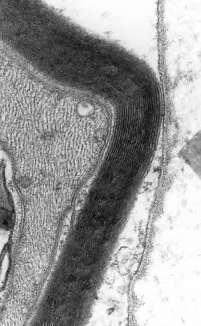
Fig. 3: Lamina over the surface of a Schwann cell: note how it follows the contour of the cell surface, in contrast to the fibronectin in figures 1 and 2.