
Vol. 35, n.º 4, 2002
|
REVISTA
ESPAÑOLA DE
Vol. 35, n.º 4, 2002 |
José Antonio Aramburu González, Ana María Dotor de Lama, Carlos Santonja
Department of Pathology. Hospital Universitario de Getafe, Madrid. Carretera de Toledo, km 12,500 (28905 Getafe, Madrid; Spain).
SUMMARY
We report a case of intracranial solitary fibrous tumour (SFT) arising on the meninges of a 51 year-old woman who also had a synchronous meningioma, an association not previously reported. SFT of the meninges showed histological findings similar to SFT in other locations and immunohistochemical study showed diffuse positive staining for CD-34 and focal staining for bcl-2. In the central nervous system this kind of tumour should be distinguished from meningiomas and haemangiopericytomas. We discuss the differential diagnosis and review the literature published about these neoplasms.
Keywords: Solitary fibrous tumour, meningioma, haemangiopericytoma, CD-34.
RESUMEN
Presentamos el caso de una mujer de 51 años con un tumor fibroso solitario meníngeo en la región occipital izquierda, que simultáneamente asociaba un meningioma convencional en la región frontal derecha. Dicha asociación no ha sido descrita anteriormente. El TFS meníngeo muestra hallazgos histológicos similares a los TFS de otras localizaciones. El estudio inmunohistoquímico muestra positividad difusa para CD-34 y focal para bcl-2. En el sistema nervioso central, el diagnóstico diferencial de estos tumores debe hacerse con el meningioma y el hemangiopericitoma. En este artículo se discute el diagnóstico diferencial y se hace una revisión de la literatura.
Palabras clave: Tumor fibroso solitario, meningioma, hemangiopericitoma, CD-34.
INTRODUCTION
The so-called solitary fibrous tumour is a relatively uncommon mesenchymal neoplasm. Although originally described in the pleura, it has recently been documented at other sites, including the nervous system.
To the best of our knowledge, only twenty examples of this tumour have been reported in intracranial localization (table I)
We report a superficially located, intracranial solitary fibrous tumour arising from the meninges of a 51 year-old woman, who also had a synchronous classic right frontal meningioma.
CLINICAL CASE
A 51 year-old woman presented with a one-year history of intense headache, gait imbalance, mental decline and vomiting. Her past medical history was unremarkable.
Neurologic examination revealed bilateral papilledema with right homonymous hemianopsia. No motor or sensory involvement was detected.
Neuroimaging demonstrated 3 extraaxial, dural-based masses: in the left parasagittal occipital region (6 cm in diameter), the right parasagittal frontal region (of unspecified size) and the left parasagittal region (1 cm in diameter). They were isointense with homogeneous intensity. The first had prominent vascularity, in contradiction with the other two lesions.
The first two tumours were totally resected. There has been no recurrence on a 3-year follow-up.
PATHOLOGIC FINDINGS
1. Left occipital tumour: Grossly, multiple firm fragments of tissue were received, measuring in aggregate 10.0 ¥ 10.0 ¥ 3.0 cm. with a homogeneous brown-grey cut surface. A fragment of duramater was also received. Histologically, this tumour was characterised by a cellular spindle-cell proliferation arranged haphazardly or in short fascicles (fig. 1), in a rich capillary network. The latter sometimes exhibited a staghorn pattern mimicking haemangiopericytoma (fig. 2), with a moderately collagenous background. The neoplastic cells displayed vesicular nuclei, finely dispersed chromatin, and inconspicuous nucleoli. Their cytoplasm was scant, poorly defined, and did not stain with PAS. The mitotic count was low (less than 3 mitoses per 10 HPF). A reticulin stain demonstrated strong pericellular positivity (fig. 3B). On immunohistochemical staining, the tumour cells exhibited strong and diffuse positive staining for vimentin (monoclonal, 1:160; microwave antigen retrieval, Dako), CD34 (monoclonal clon QBEnd/10, 1:25; Novocastra) (figure 3A) and bcl-2 protein (monoclonal, 1:160; microwave antigen retrieval, Dako). It was negative for epithelial membrane antigen (EMA), low and high molecular weight cytokeratins (AE1-AE3, CAM 5.2), S-100 protein, gliofibrillary acid protein (GFAP), muscle markers, CD10 and hormone receptors (ER, PR). The Mib-1 labelling index was less than 5%, and rare scattered cells were positive for p53 protein. Although the initial diagnostic impression was haemangiopericytoma, the final diagnosis was Solitary Fibrous Tumour (see discussion).
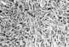
Fig. 1. A cellular region composed
of elongated or oval cells with plump nuclei arranged in short fascicles (HE x
250).
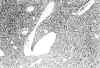
Fig. 2. Thin-walled vascular
channel with a haemangiopercytomatous pattern (HE x100).
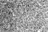
Fig. 3a. Strong and diffuse CD-34
immunoreactivity (CD-34 x250).
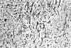
Fig. 3b. Extensive intercellular
reticular deposition (Reticulin x100).
2. Right frontal tumour: Grossly, the specimen consisted of multiple firm fragments of tumour tissue measuring in aggregate 5.0 ¥ 4.0 ¥ 1.5 cm, with a homogeneous brown-grey cut surface. Histologically, it showed a typical pattern of menigothelial cells with syncitial whorls. On immunostaining, the tumour cells were positive for vimentin, EMA and S-100 protein, and negative for CD34. The Mib-1 labelling index was less 2%. The diagnosis was Meningothelial Meningioma.
DISCUSSION
The solitary fibrous tumour (SFT) is a relatively rare neoplasm that arises most frequently in association with the pleura. To our knowledge, there are only 20 previously reported cases of intracranial SFT of the meninges (1-12). In the most recent WHO classification of neoplasms of the nervous system (Lyon 2000), SFT is accepted as a clinicopathologic entity, and it is included with other mesenchymal, non-meningothelial cell tumours. It usually occurs in adult patients, with only two cases reported in youngsters (9,11). One case had a previous history of irradiation (11) In our case the patient was an adult female with an unremarkable past medical history.
The histopathologic differential diagnosis of SFT concerns other spindle cell neoplasms that arise in the meninges. The distinction from fibroblastic meningioma is the most important and problematic (2). SFT does not show any of the typical cellular patterns of meningiomas, although fibroblastic meningioma (FM) often lacks many of the typical histological features of meningioma. SFT shows a prominent reticulin network, which is not a feature of meningiomas. Unlike meningiomas, the cytoplasm of cells of SFTs does not stain with PAS. Strong and diffuse immunohistochemical positivity for vimentin and CD34 with negative staining for EMA and S-100 protein are characteristic. However, in many previous reports SFT has been initially misinterpreted as FM (2,4,5,11-13).
Discrimination of SFT from haemangiopericytoma (HMP) is especially relevant because it carries significant therapeutic and prognostic implications. A haemangiopericytomatous pattern is present in a wide variety of neoplasms, including SFT. The staghorn vascular pattern is usually more extensive and widespread in haemangiopericytomas than in SFT (2). Immunostaining for CD34 can be focally and weakly positive in HMP (2-5,10,11); Perry (13) observed focal and weak CD34 expression in only 33% of his cases of haemangiopericytoma, in contrast to strong positivity of 100% of his cases of SFT. Brunnemann (1) claims that CD34 cannot be used to distinguish reliably SFT from HP and, since lack of immunoreactivity for CD34 does not exclude the diagnosis of SFT, he believes that SFT and HMP are closely related, perhaps even the same entity. Our case was initially interpreted as HP, but its non-aggressive behaviour (three years without tumour recurrence), as well as the intense and diffuse CD34 immunostaining prompted a diagnostic reinterpretation. Bcl-2 oncoprotein is expressed by many benign and malignant spindle cell tumours (14). SFTs, like HP, are usually positive for bcl-2 (14), but meningiomas are bcl-2 negative. Our case expressed this antigen in a very intense and diffuse manner. Immunostaining for Factor XIIIa, CD57 and Collagen IV is non contributory for diagnostic purposes (2,13).
Finally, the lack of S-100 protein immunostaining excludes a Schwannoma from the differential diagnosis.
The metastatic potential of the intracranial leptomeningeal SFT appears to be low, but tumour recurrences are common whenever total resection is not achieved (4,5,12). Only in one case has meningeal SFT metastasized outside the nervous system (5). The low MIB-1 index observed in our case and in most of the previously reported cases further supports the notion that this tumour is a slow growing neoplasm, behaving like a grade I meningioma.
Lastly, even though meningiomas can present synchronously with other oestrogen-dependent tumours, as well as be intermingled with glial tumours, we have not found previous reports of the simultaneous occurrence of SFT and meningioma.
REFERENCES
Brunnemann RB, Ro JY, G. Ordoñez N, Mooney J, El-Naggar AK, Ayala AG. Extrapleural solitary fibrous tumor: a clinicopathologic study of 24 cases. Mod. Pathol 1999; 12: 1034-42.
Carneiro Siderlei, Scheithauer BW, Nascimento AG, Hirose T, Davis DH. Solitary fibrous tumor of the meninges: a lesion distinct from fibrous meningioma. A clinicopathologic and immunohistochemical study. Am J Clin Pathol 1996; 106: 217-24.
Challa VR, Kilpatrick SE, Ricci P, Wilson JA, Kelly DL. Solitary fibrous tumour of the meninges. Clin Neuropath 1998; 17: 73-8.
Gentil Perret A., Mosnier JF, Duthel R, Brunon J, Barral F, Boucheron S. Tumeur fibreuse solitaire des méninges. Ann Pathol 1999; 19: 532-5.
Ng HK, Choi PC, Wong CW, To KF, Poon WS. Related Articles. Metastatic solitary fibrous tumor of the meninges. Case report. J Neurosurg 2000; 93:490-3.
Nawashiro H, Nagakawa S, Osada H, Katoh H, Ohnuki A, Tsuzuki N. Solitary fibrous tumor of the meninges in the posterior cranial fossa: magnetic resonance imaging and histological correlation. A case report. Neurol. Med Chir (Tokio) 2000; 40: 432-4.
Nikas DC, De Girolami U, Folkerth RD, Bello L, Zamani AA, Black PM. Parasagittal solitary fibrous tumour of the meninges. Case report and review of the literature. Acta Neurochir Wien 1999; 141: 307-3.
Prayson RA, McMahon JT, Barnett GH. Solitary fibrous tumor of the meninges. Case report and review of the literature. J Neurosurg 1997; 86: 1049-52.
Rodríguez L, López J, Marín A, Cardozo D, Molina O, Cardozo J. Solitary fibrous tumor of the meninges. Clin Neuropath 2000; 19: 45-8.
Shimizu S, Oka H, Kawano N, Utsuki S, Suzuki S, Iwabuchi K, Kan Fujii K. Solitary fibrous tumor arising from the falx cerebri. Case report. Neurol Med Chir (Tokio) 2000; 40: 650-4.
Slavik T, Bentley RC, Gray L, Fuchs HE, Mclendon RE. Solitary fibrous tumor of the meninges occurring after irradiation of a mixed germ cell tumor of the pineal gland. Clin Neuropathol 1998; 17: 55-60.
Suzuki SO, Fukui M, Nishio S, Iwaki T. Clinicopathological features of solitary fibrous tumor of the meninges: An immunohistochemical reappraisal of cases previously diagnosed to be fibrous meningioma or hemangiopericytoma. Pathol Int 2000; 50: 808-17.
Perry A, Scheithauer BW, Nascimento AG. The immunophenotypic spectrum of meningeal hemangiopericytoma: A comparison with fibrous meningioma and solitary fibrous tumor of meninges. Am J Surg Pathol 1997; 21: 1354-60.
Miettinen M, Sarlomo-Rikala M, Kovatich AJ. Cell-type and tumor-type related patterns of bcl-2 reactivity in mesenchymal cells and soft tissue tumors. Virchows Arch 1998; 433: 255-60.
Acknowledgements: We would like to acknowledge the help of María del Mar Granados Alamillo in performing the immunohistochemical stains. José Domínguez kindly digitalized the microscopic photographs.
![]()